CSBWFU Facilities
X-Ray Diffraction Laboratory
The X-Ray Diffraction Laboratory
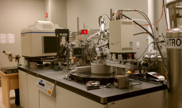 consists of a Rigaku/MSC MicroMax-007 Microfocus
rotating anode generator, Saturn-92 Digital CCD Camera system, and an R-AXIS IV imaging plate system,
which was upgraded with an inverse phi axis. Both ports on the generator are outfitted
with Varimax HR optics.
The protein crystallization facilities are conveniently located adjacent to the
X-Ray Diffraction Laboratory; these include a 100 ft2 cold room
dedicated to crystallization, a Belle Technology Clear View Glove Box for
consists of a Rigaku/MSC MicroMax-007 Microfocus
rotating anode generator, Saturn-92 Digital CCD Camera system, and an R-AXIS IV imaging plate system,
which was upgraded with an inverse phi axis. Both ports on the generator are outfitted
with Varimax HR optics.
The protein crystallization facilities are conveniently located adjacent to the
X-Ray Diffraction Laboratory; these include a 100 ft2 cold room
dedicated to crystallization, a Belle Technology Clear View Glove Box for
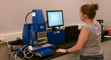 preparing and cryo-cooling crystals in the absence of oxygen, and a Gryphon high throughput crystallization robot.
Grants from the North Carolina Biotechnology Center funded the purchase of the Gryphon robot, in addition to a 4DX Systems AB single-crystal microspectrophotometer and a Tecan Safire2 monochromator-based microplate reader.
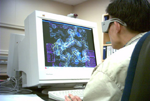
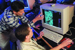 includes 9 Linux workstations utilizing hardware stereo for structural
biology applications. E-mail and web services are handled through an internally managed departmental server.
Two additional servers function as the Computational Biology Resource for the
Biomolecular Resource Facility and in file hosting operations, respectively.
includes 9 Linux workstations utilizing hardware stereo for structural
biology applications. E-mail and web services are handled through an internally managed departmental server.
Two additional servers function as the Computational Biology Resource for the
Biomolecular Resource Facility and in file hosting operations, respectively.
 The BCGF also includes a 16-node computational Linux cluster with the latest
software for high performance applications such as macromolecular structure determination,
molecular modeling, and structure based drug design.
In addition, the
The BCGF also includes a 16-node computational Linux cluster with the latest
software for high performance applications such as macromolecular structure determination,
molecular modeling, and structure based drug design.
In addition, the